
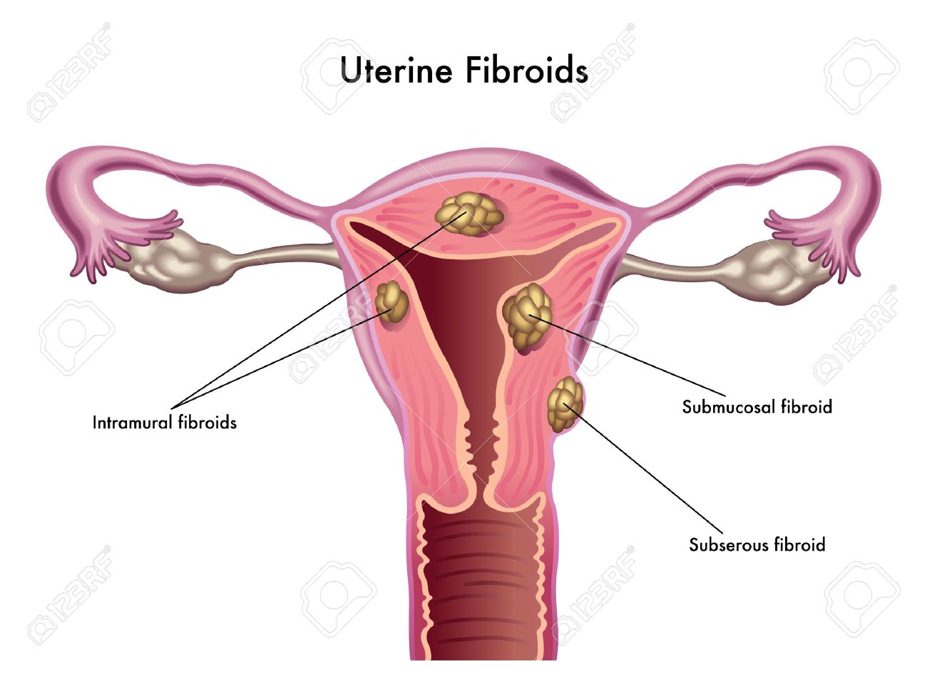
فیبروم رحم رشد غیرسرطانی سلول ها است که در دیواره عضلانی رحم ایجاد می شود. فیبروم اندازه های مختلفی می تواند داشته باشد. گاهی کمتر از یک سانتی متر و گاهی نیز آنقدر بزرگ است که باعث رشد رحم به اندازه ماه پنجم بارداری می شود. فیبروم به تنهایی باعث ایجاد علامتی نمی شود، اما اندازه و محل قرارگیری آن ها می تواند خونریزی شدید و درد زیر شکم ایجاد کند. بین 80 تا 90 درصد خانم های مبتلا به فیبروم احتیاجی به درمان پیدا نمی کنند. فیبروم رحم با معاینه متخصص زنان و سونوگرافی تشخیص داده می شود. فیبروم رحم گاهی نیز با نام میوم رحم شناخته می شود. فیبروم از نطر محل قرارگیری آن انواع مختلفی دارد.
علامت های فیبروم رحم
- پریود طولانی و همراه با درد شدید؛ همچنین خونریزی غیرمعمول واژینال همراه با لخته های خون. این می تواند باعث کم خونی در شخص شود.
- فشار و درد زیر شکم
- کمر درد و پا درد
- درد در زمان رابطه جنسی
- فشار آمدن به مثانه و تکرر ادرار
- فشار آمدن به روده، یبوست و نفخ
- بزرگ شدن شکم به طور غیر معمول
فیبروم در خانم های باردار مشکلی برای جنین ایجاد می کند؟
خوشبختانه در اغلب موارد فیبروم تداخلی با بارداری ندارد؛ اما ممکن است لوله های فالوپ را تحت تأثیر قرار دهد و برای باردار شدن شخص در آینده مشکل ایجاد کند. البته به ندرت فیبروم باعث ایجاد خطر سقط جنین نیز می شود.
با بارداری شخص فیبروم به تدریج بزرگ تر می شود. این به خاطر افزایش سطح استروژن در زمان بارداری است. بعد از زایمان فیبروم دوباره به اندازه اولیه خود بر می گردد. بعد از یائسگی یعنی زمانی که هورمون استروژن در بدن افت می کند، فیبروم نیز بهبود می یابد. البته در صورت مصرف مکمل دارویی استروژن علائم فیبروم بهبودی پیدا نمی کند. بعضی از انواع فیبروم ها برای لانه گزینی و رشد جنین مشکلاتی را ایجاد می کنند. در این صورت قبل از بارداری بهتر است فیبروم درمان شود.
درمان فیبروم
برای درمان فیبروم روش های مختلفی به کار می رود. این روش ها شامل تکنیک های جراحی و غیرجراحی و نیز مصرف دارو هستند. با مصرف داروهایی که توسط پزشک تجویز می شوند می توان از رشد فیبروم جلوگیری کرد یا آن را کند نمود.
به طور کلی تکنیک هایی که تا کنون برای درمان فیبروم رحم توسعه پیدا کرده اند شامل موارد زیر است:
هیسترکتومی
هیسترکتومی به معنی جراحی برداشتن رحم است. هیسترکتومی می تواند به دو روش جراحی باز یا لاپاراسکوپی انجام شود. جراحی باز به چهار روز بستری در بیمارستان نیاز دارد و بعد از ترخیص نیز بیمار باید شش هفته در منزل استراحت کند. لاپاراسکوپی تکنیکی با حداقل تهاجم است و بیمار بعد از آن احساس ناراحتی کمتری می کند.
میومکتومی
میومکتومی روشی برای درمان فیبروم است که بسته به اندازه و محل ایجاد فیبروم، ممکن است به صورت سرپایی و یا همراه با دو یا سه روز بستری در بیمارستان انجام شود. در روش میومکتومی فیبروم یا فیبروم های روی رحم برداشته و رحم دوباره دوخته می شود. میومکتومی این مزیت را دارد که رحم باقی می ماند و شخص می تواند به حفظ باروری و توانایی باردار شدن در آینده امیدوار باشد.
كرايو سرجري
این تکنیک می تواند به صورت لاپاراسکوپی، واژینال و یا زیرپوستی (percutaneously) انجام شود. این روش روشی کم تهاجم، امن و مؤثر برای درمان و بهبود علائم فیبروم رحم است. این روش برای خانم هایی مناسب است که می خواهند رحمشان سالم و دست نخورده بماند اما تمایل به بارداری در آینده ندارند.
آمبولیزاسیون شریان رحمی
آمبولیزاسیون شریان رحمی در بیمارانی که تمایل به انجام جراحی ندارند بسیار مفید است. در این تکنیک جریان خونی که فیبروم را تغذیه می کند متوقف می شود و به این ترتیب فیبروم به مرور کوچک می شود و از بین می رود.
استفاده از امواج اولتراسوند تحت هدايت ام آر آي
استفاده از امواج اولتراسوند تحت هدايت ام آر آي تکنیکی جدید با حداقل تهاجم برای درمان فیبروم است که بدون ایجاد برشی روی بدن انجام می شود. این روش تلفیقی از پرتو اولتراسوند برای ایجاد حرارت و تخریب بافت بیمار و استفاده از سیستم تصویربرداری ام آر آی برای مشاهده داخل بدن بیمار است.
از بین روش های ذکر شده روش هیسترکتومی و میومکتومی روش هایی مرسوم برای درمان فیبروم رحم هستند. متخصصان حوزه زنان و زایمان موفقیت این دو روش در طول سال ها ثبت کرده اند.
دكتر حقگو برای رعایت حال بیماران و تضمین سلامتی خانم ها انواع جراحي های فيبروم رحم را بدون باز نمودن شكم و با روش لاپاروسكوپي و هيستروسكوپي انجام مي دهند.
منابع
http://www.sirweb.org//
http://www.medicinenet.com/
http://www.medicinenet.com/
http://www.mayoclinic.org/
http://www.thebump.com/
http://www.fudahospital.com/
http://www.insightec.com/
Uterine fibroids are very common non-cancerous (benign) growths that develop in the muscular wall of the uterus. They can range in size from very tiny (a quarter of an inch) to larger than a cantaloupe. Occasionally, they can cause the uterus to grow to the size of a five-month pregnancy. In most cases, there is more than one fibroid in the uterus. While fibroids do not always cause symptoms, their size and location can lead to problems for some women, including pain and heavy bleeding.
Uterine fibroids are the most common tumors of the female genital tract. You might hear them referred to as “fibroids” or by several other names, including leiomyoma, leiomyomata, myoma and fibromyoma. Fibroid tumors of the uterus are very common, but for most women, they either do not cause symptoms or cause only minor symptoms. Most fibroids don’t cause symptoms—only 10 to 20 percent of women who have fibroids require treatment.
Uterine Fibroid Symptoms
Depending on size, location and number of fibroids, they may cause:
Heavy, prolonged menstrual periods and unusual monthly bleeding, sometimes with clots; this can lead to anemia
- Pelvic pain and pressure
- Pain in the back and legs
- Pain during sexual intercourse
- Bladder pressure leading to a frequent urge to urinate
- Pressure on the bowel, leading to constipation and bloating
- Abnormally enlarged abdomen
How will uterine fibroids affect my baby?
Luckily, they usually don’t interfere with pregnancy. It’s possible that uterine fibroids can change the shape of or block your fallopian tubes, which can affect future pregnancies. In some cases, doctors may recommend removing problematic fibroids before you get pregnant. You might experience some pain in your lower abdomen — if you do, ask your doctor what you should do. Most of the time, you can take medication for the pain. In some cases, uterine fibroids can increase the risk of miscarriage, preterm birth or breech birth. You could also have a greater chance of getting a c-section or heavy bleeding after labor.
Fibroids can dramatically increase in size during pregnancy. This is thought to occur because of the increase in estrogen levels during pregnancy. After pregnancy, the fibroids usually shrink back to their pre-pregnancy size. They typically improve after menopause when the level of estrogen, the female hormone that circulates in the blood, decreases dramatically. However, menopausal women who are taking supplemental estrogen (hormone replacement therapy) may not experience relief of symptoms.
Fibroids usually don’t interfere with conception and pregnancy. However, it’s possible that fibroids could cause infertility or pregnancy loss. Submucosal fibroids may prevent implantation and growth of an embryo. In such cases, doctors often recommend removing these fibroids before attempting pregnancy or if you’ve had multiple miscarriages. Rarely, fibroids can distort or block your fallopian tubes, or interfere with the passage of sperm from your cervix to your fallopian tubes.
What is the treatment for uterine fibroids?
There are several options for the treatment of uterine fibroids that include surgery (hysterectomy, myomectomy, cryosurgery, MRI-guided high-intensity focused ultrasound (MRgFUS), and uterine artery embolization (UAE). edical treatments include medications such as mifepristone (RU-486, danazol (Danocrine), raloxifene (Evista), GnRH analogs (Lupron and others), and low-dose formulations of oral contraceptives. Surgical treatments for fibroids are briefly descrived below:
Hysterectomy
Hysterectomy is the removal of the uterus and is considered major abdominal surgery. It requires three to four days of hospitalization and the average recovery period is six weeks. The newer and more sophisticated procedures use laparoscopy to assist the hysterectomy procedure.
Myomectomy
Depending on the size and placement of the fibroids, myomectomy can be an outpatient surgery or require two to three days in the hospital. However, myomectomy is usually major surgery that involves cutting out the biggest fibroid or collection of fibroids and then stitching the uterus back together. Myomectomy leaves the uterus in place and may, therefore, preserve the woman’s ability to have children.
Cryomyolysis
Cryomyolysis which is performed by laparoscopically, transvaginally or percutaneously, is a minimally invasive, safe, and feasible procedure for obtaining myoma shrinkage and symptom relief in women with symptomatic fibroids who wish to preserve their uterus but do not desire future pregnancies.
Uterine artery embolization
In appropriately selected patients, uterine artery embolization (UAE) can be an effective treatment option, especially for women who for a variety of reasons are not good candidates for surgery. This reduces or stops blood flow to the fibroids, which in turn causes them to die and shrink.
MR guided Focused Ultrasound Surgery (MRgFUS) technology
MR guided Focused Ultrasound Surgery (MRgFUS) technology is a new treatment choice without knife or needle. MRgFUS is a completely non-invasive treatment option for treating fibroids. It combines a high intensity focused ultrasound beam that heats and destroys targeted tissue, non-invasively and Magnetic Resonance Imaging system (MRI) which visualizes patient anatomy, and controls the treatment by continuously monitoring the tissue effect.
References
http://www.sirweb.org//
http://www.medicinenet.com/
http://www.medicinenet.com/
http://www.mayoclinic.org/
http://www.thebump.com/
http://www.fudahospital.com/
http://www.insightec.com/